
By Dr. Myriah Hinchey, ND, FMAPS, FILADS, Medical Director, TAO Vitality | Founder, LymeCore Botanicals
Summary: The acute window following a tick bite is the highest-yield period for early detection and also the period when standard diagnostic approaches are least reliable. A sound tick bite testing protocol for this window must account for the biological factors that limit standard testing and prioritize the methods best matched to the timing of exposure. This article, adapted from a Galaxy Diagnostics clinical education webinar presented by Dr. Myriah Hinchey, ND, FMAPS, FILADS, outlines the biological and methodological factors that limit standard testing in the acute phase, explains the distinction between direct and indirect detection, and provides a practical framework for co-infection-aware diagnostic decision-making in the days and weeks following a known or suspected tick bite.
Table of Contents
The acute period following a tick bite is one of the most consequential and most diagnostically constrained windows in tick-borne disease medicine. Standard testing is unreliable this early. Antibodies have not yet formed. Symptoms may be nonspecific or absent. And the organisms transmitted by a single bite often carry timelines and presentations that clinicians underestimate.
What Is a Tick Bite Testing Protocol?
A tick bite testing protocol is a structured, timing-aware approach to pathogen detection following known or suspected tick exposure. It accounts for the antibody lag window that limits standard serology in early infection, prioritizes direct detection methods during the acute phase, and evaluates for co-infecting organisms simultaneously rather than sequentially. The framework described in this article is adapted from a Galaxy Diagnostics clinical education webinar and reflects current evidence on transmission timing, pathogen biology, and detection methodology.
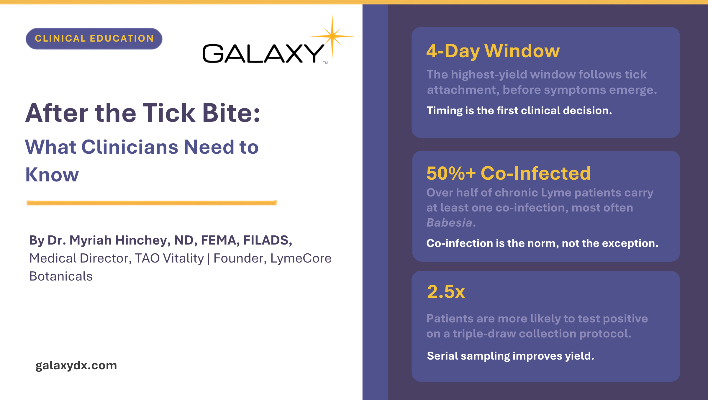
The 4-Day Critical Window: Why Timing Is the First Clinical Decision
Borrelia burgdorferi, the causative organism of Lyme disease, resides in the hind gut of the tick during quiescence. When the tick begins a blood meal, the presence of blood in the tick’s intestines mobilizes the spirochetes, which migrate to the salivary glands and can then be injected into the host. This process takes time, which is why prompt tick removal matters. However, clinicians should be aware that partially-fed ticks, those that have already taken a partial blood meal from another animal or host, may already have spirochetes positioned in the salivary glands and can transmit Borrelia more rapidly upon reattachment [1].
Other co-infecting pathogens operate on shorter transmission timelines. Rickettsia species and Powassan virus can both be transmitted rapidly – in the case of Powassan, within as little as 15 minutes of attachment [2,3]. By the time a clinician sees a patient who has found an attached tick, transmission of certain organisms may have already occurred, regardless of how quickly the tick was removed.
If Borrelia infection goes unrecognized and unaddressed, spirochetes can disseminate into the cerebrospinal fluid within weeks of initial infection [4]. Multi-system involvement follows, with inflammatory, neurological, cardiac, and joint sequelae. Chronic tick-borne disease is significantly more difficult to address clinically than early infection. The biological case for detection in the acute window is direct.
What This Means for Testing
A tick bite testing protocol in the acute phase should prioritize direct detection and should not use a negative serological result to exclude infection. The antibody lag window renders standard serological testing diagnostically unreliable in the first 3 to 6 weeks post-exposure. Direct detection methods, those that detect the pathogen’s own genetic material or antigens rather than the host’s antibody response, are better suited to this period. Baseline laboratory monitoring with CBC and liver function tests can provide early signals for co-infections such as Anaplasma and Ehrlichia, which produce characteristic hematologic changes.
Clinical Presentation: Why Lyme Disease and Co-Infections Are Frequently Missed
Lyme disease and the co-infections transmitted by the same ticks present with overlapping, nonspecific symptoms that do not consistently point toward a single diagnosis. Clinicians who see these patients in the acute phase often encounter a symptom picture that mimics viral illness, allergic response, autoimmune conditions, or other common diagnoses. This is not incidental – it is a consistent pattern documented across clinical series.
Borrelia infection progresses in stages. Early localized disease presents with flu-like symptoms including fatigue, fever, chills, headache, muscle aches, and possible neck pain, with or without an erythema migrans (EM) rash. Early disseminated disease can involve Lyme carditis, lymphocytoma, facial palsy, joint stiffness, and Lyme neuroborreliosis. Late or chronic disseminated disease presents with persistent, cyclic, multi-system symptoms, severe arthritis typically involving one knee, and neuropsychiatric manifestations attributable to the pathogen’s capacity to cross the blood-brain barrier.
Co-infections complicate this presentation substantially. A patient carrying multiple tick-borne pathogens simultaneously will present with overlapping and compounding symptoms that can shift the entire clinical picture. A survey of more than 3,000 patients with chronic Lyme disease found that over 50% had at least one co-infection and 30% had two or more [5]. Babesia was the most frequently identified co-infection, followed closely by Bartonella.
Each co-infecting organism has its own characteristic onset timing and clinical signature:
Pathogen | Typical Onset | Characteristic Findings |
Babesia | 1 to 4 weeks | Air hunger, hemolytic anemia, fever, night sweats, fatigue, jaundice, dark urine, thrombocytopenia |
Bartonella | Weeks to months | Profound headache, lymphadenopathy, striae or pustular rash, encephalopathy, neuropsychiatric symptoms |
Anaplasma / Ehrlichia | 1 to 2 weeks | High fever, severe headache, leukopenia, thrombocytopenia, elevated liver enzymes |
Rickettsia | Within 1 week | Sudden high fever, petechial or maculopapular rash, eschar at bite site, urgent, potentially life-threatening |
Mycoplasma | 1 to 3 weeks | Fever, cough, headache, fatigue, muscle stiffness, chest pain |
Powassan virus | 1 to 5 weeks | Encephalitis, seizures, confusion, no available antiviral; early recognition critical |
These co-infections are frequently misattributed to other diagnoses when clinicians encounter them without a confirmed tick exposure history. Babesia is commonly misdiagnosed as anemia, malaria, heart failure, lupus, or MS. Bartonella is frequently misattributed to schizophrenia, ADHD, OCD, autism, or depression, particularly in pediatric patients. Borrelia itself is misdiagnosed as Alzheimer’s disease, ALS, heart failure, or rheumatoid arthritis in chronic presentations.
The more co-infecting pathogens are present, the more synergistically they contribute to immune dysregulation and inflammation. Multi-pathogen infections are generally associated with more severe illness, broader symptom distribution, and more complex and prolonged clinical courses [6].
What This Means for Testing
Co-infection testing tick bite workups should begin at the first clinical encounter, not after a single-pathogen result returns. A single-pathogen testing approach is not appropriate for tick-borne disease in either the acute or chronic setting. The co-infection prevalence data, combined with overlapping symptom patterns, supports a comprehensive testing strategy that evaluates Borrelia, Babesia, and Bartonella simultaneously, along with Anaplasma, Ehrlichia, and Rickettsia where clinical or epidemiological context warrants. Standard CBC and liver function tests provide early signal for Anaplasma, Ehrlichia, and Rickettsia and should not be overlooked in the acute workup. Because Rickettsia (Rocky Mountain spotted fever) and Anaplasma/Ehrlichia can be life-threatening, any combination of sudden high fever, severe headache, and hematologic abnormalities in the context of tick exposure warrants urgent clinical attention.
The EM Rash: Diagnostic Significance and Its Limits in Practice
The classic bullseye presentation, concentric rings radiating from the bite site, appears in fewer than 20% of Lyme disease cases, with some published studies reporting rates as low as 6% [7]. The more common presentation is a solid pink or red area that can be mistaken for an insect bite, contact dermatitis, or cellulitis. A vesicular or blistering rash at or near the bite site may suggest Bartonella co-infection and warrants evaluation accordingly. Approximately 20-30% of patients with confirmed Lyme disease develop no rash at all [8]
Clinicians should also note that EM rashes are typically asymptomatic at the skin surface. They are often not warm, tender, or raised in a way that would independently prompt a patient to seek care. This contributes to cases being missed or dismissed until the infection has disseminated.
What This Means for Testing
When an EM rash is observed, the diagnostic question is no longer whether Lyme disease is present, it is what co-infections may also be present. Testing should focus on co-infection evaluation and baseline laboratory monitoring rather than on confirming the Borrelia diagnosis the rash already provides. A negative serological result in this context does not change the clinical picture. Direct detection methods can still be used to document organism presence in the submitted sample or to evaluate for co-infecting pathogens where clinical concern exists.
Why Standard Lyme Disease Testing After a Tick Bite Is Structurally Limited in the Acute Phase
The standard approach for Lyme disease testing in most clinical settings is the CDC-recommended two-tier algorithm: an initial ELISA, followed by a Western blot if the ELISA is positive or equivocal. This approach is serological, it detects the host antibody response to Borrelia, not the organism itself.
The structural limitations of this approach in the acute setting are well-documented. One analysis found that up to 75% of Lyme disease cases were missed using CDC-recommended methodology [9]. A separate review reported a detection rate of approximately 54% across confirmed cases [10]. Part of this gap is attributable to antibody timing. IgM antibodies peak at approximately 3 weeks post-infection. IgG antibodies peak around 6 weeks [11]. Testing before these windows have elapsed produces negative results in patients with confirmed early infection, not because the test failed technically, but because the biology has not yet generated what the test is designed to detect.
Strain coverage represents an additional structural gap. The standard two-tier assay is designed to detect Borrelia burgdorferi sensu stricto. It does not reliably detect the multiple B. burgdorferi sensu lato genospecies now recognized as causative agents of Lyme borreliosis across North America and Europe, nor does it address tick-borne relapsing fever caused by other Borrelia species [12] A negative result on the standard two-tier algorithm cannot rule out Lyme disease attributable to these additional species.
Perhaps the most significant gap in co-infection terms is that the two-tier algorithm does not test for Babesia, Bartonella, Ehrlichia, Anaplasma, Rickettsia, Mycoplasma, or Powassan virus. A clinician who orders standard Lyme serology and receives a negative result has not tested for any of the co-infections that the same tick may have transmitted simultaneously. These are separate clinical questions requiring separate testing.
What This Means for Testing
A negative standard two-tier test in the acute phase should be recognized as an expected methodological result, not as evidence that infection is absent. Acute Lyme disease testing with standard serology cannot answer whether infection is present during the first 3 to 6 weeks post-exposure, the biology and timing do not allow it to produce meaningful information in that window. Specialty laboratory testing using direct detection methods and broader species coverage provides more actionable information during this period. Standard testing may have a defined role in follow-up evaluation at 3 to 6 weeks post-exposure, when antibody titers are more interpretable.
Direct vs. Indirect Detection: The Diagnostic Foundation of a Tick Bite Testing Protocol
Understanding the distinction between direct and indirect testing is foundational to interpreting results accurately and selecting the right method at the right time. These categories measure fundamentally different biological signals, and no single test provides the complete diagnostic picture.
Direct testing detects the pathogen itself: its DNA (via PCR or digital PCR), its RNA (via FISH), or specific antigens it sheds into host tissue or urine. Because direct testing does not depend on the host immune response, it can detect infection during the acute phase, before antibodies have formed. A positive direct result confirms that the pathogen’s material was present in the submitted sample at the time of collection.
Indirect testing detects the host immune response to the pathogen, specifically the IgG and IgM antibodies produced when the immune system encounters the organism. A positive indirect result indicates that the patient’s immune system has responded to the pathogen. It does not, on its own, distinguish between current pathogen presence and prior exposure. Indirect testing is most interpretable once antibody titers have reached detectable thresholds, which in tick-borne disease is generally 3 to 6 weeks after infection.
Direct Testing | Indirect Testing | |
What it detects | Pathogen DNA, RNA, or antigen | Host antibody response (IgG, IgM) |
Examples | PCR, dPCR, FISH, urine antigen | ELISA, Western blot, IFA, immunoblot |
Optimal use window | Acute phase | 3 to 6 weeks post-exposure |
Key strength | Detects before antibodies form | Widely available; broadly applicable |
Key limitation | Pathogen abundance may be low; sampling variability | Cannot detect in acute window; does not confirm current pathogen presence |
A useful framework for thinking about direct testing is sampling probability. A direct test can only identify what is present in the blood collected at the time of the draw. Tick-borne pathogens circulate at low abundance and may not be present in detectable concentrations in every blood draw. A negative direct test result means the organism was not detected in the submitted sample at that moment. It does not confirm that the organism is absent from the patient.
This is why Galaxy Diagnostics recommends a three-day serial draw protocol for its digital PCR testing. Patients are approximately 2.5 times more likely to have a detected result on a triple-draw collection than on a single draw [13]. In cases where pathogen DNA was detected across a triple draw, it was frequently positive on only one of the three draws, underscoring the variability of low-abundance organisms in circulation.
Digital PCR (dPCR) improves sensitivity over standard quantitative PCR (qPCR) by partitioning each blood sample into thousands of individual reaction chambers. Each partition is read independently as positive or negative, allowing absolute quantification of target DNA even at concentrations that fall below the detection threshold of standard qPCR. In the context of acute tick-borne disease, where pathogen burden in blood may be low, this sensitivity difference is clinically meaningful.
What This Means for Testing
In a tick bite testing protocol, direct vs. indirect Lyme testing are not interchangeable, they are complementary tools applied at different points on the clinical timeline. In the acute phase, direct detection methods are more appropriate than serological approaches because they do not depend on an immune response the patient has not yet mounted. A negative direct test result during the acute window should be interpreted within the full clinical context: the patient’s symptom pattern, timing of exposure, and whether serial sampling was used. Indirect testing has a defined role in follow-up evaluation at 3 to 6 weeks post-bite, when serological results become more interpretable.
How to Structure a Tick Bite Testing Protocol After Exposure
For clinicians managing a patient with a known or suspected tick bite, the following considerations may help structure early diagnostic decision-making. The goal is to match the testing method to the clinical question, account for the timing of the exposure, and assess for co-infection from the beginning rather than sequentially.
Clinical framing questions for a tick bite testing protocol:
- What is the timing of the exposure relative to testing? If the patient presents within the first 3 to 6 weeks of a known tick bite, serological testing is unlikely to yield actionable information. Direct detection methods should be prioritized.
- What pathogens could have been transmitted? A single bite can transmit multiple organisms simultaneously. Testing limited to Borrelia alone will miss co-infections that may be shaping the clinical presentation. Testing should reflect geographic distribution of tick species and the full range of pathogens they carry.
- Is an EM rash present? If yes, this constitutes a clinical diagnosis of Lyme disease. The testing question shifts to co-infection evaluation. A negative serological result does not alter this clinical assessment.
- Are there laboratory signals pointing to specific co-infections? Leukopenia, thrombocytopenia, and elevated liver enzymes in the context of fever and headache following tick exposure warrant urgent consideration of Anaplasma, Ehrlichia, or Rickettsia. CBC and liver function tests should be part of the early workup and repeated at intervals if symptoms evolve.
- Has a serial draw approach been considered? Given the low abundance and intermittent circulation of tick-borne pathogens, a single blood draw may not be sufficient for direct detection. Serial sampling improves detection probability in patients with low pathogen burden.
Galaxy Diagnostics’ Lyme Borrelia Direct Detect (urine antigen test) detects antigen shed by Borrelia burgdorferi, in urine. In a published study of patients with confirmed EM rash, the test demonstrated 100% sensitivity for detecting Borrelia antigen in the submitted samples [14]. It can be ordered as early as days 5 to 7 following tick exposure, making it one of the earliest-available direct detection options in the tick bite testing protocol window. For pediatric patients, the urine collection format offers a practical advantage over blood-based testing in an already stressful clinical encounter.
Galaxy’s BBB Direct Detect (dPCR) assay is designed for molecular detection of Borrelia, Bartonella, and Babesia simultaneously, using a three-day serial draw collection protocol. This combined approach addresses the low-abundance challenge of direct detection while covering the three most clinically significant co-infection genera in a single testing strategy. For clinicians working through complex acute presentations, testing these three pathogens in parallel rather than sequentially reflects how they are transmitted and how they present clinically.
Evaluating Specialty Laboratory Options
Not all specialty laboratories approach tick-borne disease testing in the same way. When evaluating a laboratory for a tick bite testing protocol, the following questions are clinically relevant:
- Has the laboratory published peer-reviewed studies validating its testing methods? Are those publications available for review?
- What is the antigen source used in serological assays, clinical isolates or recombinant antigens? What are the sensitivity and specificity implications of each for the population being tested?
- What detection technology is in use, PCR, digital PCR, FISH, culture, and what are the known sensitivity and species-coverage differences across methods? Digital PCR tick-borne disease testing offers meaningful sensitivity advantages over standard qPCR in low-pathogen-burden settings.
- Does the laboratory’s testing cover all relevant species of Borrelia, Babesia, and Bartonella, or only the most commonly referenced strains?
Galaxy Diagnostics is backed by more than 450 peer-reviewed publications and uses a disclosed, multiplexed dPCR platform with absolute quantification [15, 16]. The laboratory’s antigen sources, validation methodology, and test performance data are available to clinicians on request. Learn more at the Suspected Tick-Borne Bundle page on galaxydx.com.
Disclosure: The Lyme Borrelia Direct Detect (urine antigen test) and BBB Direct Detect (dPCR) are Galaxy Diagnostics laboratory developed tests (LDTs). This content is intended for educational purposes only and does not constitute medical advice, treatment guidance, or diagnostic guarantees.
Frequently Asked Questions
Why does standard Lyme disease testing miss so many patients in the acute phase?
Standard Lyme testing misses acute-phase infections because it measures the patient’s antibody response to Borrelia, not the organism itself. Antibody production takes 3 to 6 weeks to reach detectable levels following infection. Testing within this window will produce negative results in patients with confirmed early infection because the immune system has not yet generated what the test is designed to detect. This is not a test performance issue in the conventional sense, it is a structural limitation of indirect testing applied outside its reliable window. Published analyses have found that up to 75% of Lyme disease cases are missed using standard CDC methodology.
How quickly can tick-borne pathogens be transmitted after a tick attaches?
Some tick-borne pathogens can transmit within minutes of attachment. Rickettsia species and Powassan virus can be transmitted rapidly (Powassan within 15 minutes) of tick attachment. Borrelia transmission is generally slower, as the spirochetes must migrate from the tick’s hind gut to its salivary glands before injection can occur. However, partially-fed ticks, those that have already taken a blood meal from a previous host, may have spirochetes already present in the salivary glands and can transmit more rapidly upon reattachment. Clinicians should not reassure patients that a recently attached tick carries low risk based on estimated attachment duration alone, particularly when tick feeding history is unknown.
Does the absence of an EM rash rule out Lyme disease?
No. Approximately 20-30% of patients with confirmed Lyme disease develop no rash at all. Among those who do develop a rash, fewer than 20% present with the classic bullseye appearance most associated with the diagnosis. Most EM rashes are solid-colored and can be mistaken for an insect bite, cellulitis, or contact dermatitis. The absence of a rash should not be used to exclude Lyme disease from the clinical differential, and a negative test result does not change this position.
What is the difference between direct and indirect testing for tick-borne diseases?
Direct testing detects the pathogen itself; indirect testing detects the patient’s immune response to it. Direct testing uses methods such as PCR, dPCR, FISH, and urine antigen assays and does not depend on the patient’s immune response, making it appropriate for the acute window. Indirect testing detects IgG and IgM antibodies produced in response to the pathogen. Methods include ELISA, Western blot, IFA, and immunoblot. These are most interpretable 3 to 6 weeks after infection, once antibody titers have reached detectable levels. Understanding direct vs. indirect Lyme testing is foundational to any tick bite testing protocol: the two categories are not interchangeable and are most useful when applied at the appropriate points in the clinical timeline.
When should clinicians order co-infection testing after a tick bite?
Co-infection testing should be ordered from the initial workup, not reserved for follow-up. The same tick that transmits Borrelia can simultaneously transmit Babesia, Bartonella, Anaplasma, Ehrlichia, Rickettsia, or other pathogens. Waiting for Borrelia results before evaluating for co-infections delays assessment of potentially serious and independently treatable conditions. In the acute window, direct detection of co-infecting pathogens is preferred. Standard CBC and liver function tests can identify hematologic changes associated with Anaplasma, Ehrlichia, and Rickettsia and should be part of the early workup regardless of which specialty testing is ordered.
What does the Galaxy Lyme Borrelia Direct Detect (urine antigen test) detect, and when can it be ordered?
The Galaxy Lyme Borrelia Direct Detect (urine antigen test) detects antigen shed by Borrelia burgdorferi, directly in urine. It is a direct detection method that does not depend on the patient’s antibody response, making it appropriate for the acute window and a practical first-line option in a tick bite testing protocol. In a published study of patients with confirmed EM rash, the test demonstrated 100% sensitivity for detecting Borrelia antigen in the submitted samples. The test can be ordered as early as days 5 to 7 following tick exposure. The urine collection format makes it practical for pediatric populations where blood-based testing may present logistical challenges.
How does digital PCR differ from standard PCR for tick-borne pathogen detection?
Digital PCR (dPCR) detects pathogen DNA at concentrations too low for standard quantitative PCR (qPCR) to measure, making it better suited to the low-abundance conditions common in tick-borne disease. Both methods detect pathogen DNA, but they differ in how they handle low-abundance targets. Standard qPCR measures amplification in a single reaction and estimates target DNA relative to a calibration standard. Digital PCR partitions the sample into thousands of individual reaction chambers, each containing zero or one copy of the target DNA, reads each partition independently as positive or negative, and calculates the absolute quantity of target DNA from the count of positive partitions. In digital PCR tick-borne disease testing, this sensitivity difference is clinically meaningful: pathogen burden in blood may be very low in both the acute phase and in chronic presentations, and dPCR’s absolute quantification approach provides detection where standard qPCR would return a negative result.
Why is a negative standard Lyme disease test not sufficient to rule out infection?
A negative standard Lyme test cannot rule out infection for three reasons: the test measures antibody response that may not yet have formed in the acute window; published analyses show it misses up to 75% of confirmed cases even outside that window; and it does not evaluate non-burgdorferi Borrelia species or any co-infections. A negative result on the standard two-tier algorithm reflects that method’s performance within its structural constraints. In the acute window, it is negative because antibodies have not yet formed, not because the organism is absent. At later time points, the test has been shown to miss up to 75% of confirmed cases. The standard assay also does not evaluate Borrelia species beyond B. burgdorferi sensu stricto, and it does not evaluate co-infections at all. A negative standard Lyme test should be interpreted within the full clinical context: the patient’s symptom pattern, exposure history, the specific limitations of the test at the time it was ordered, and what testing has and has not yet been performed.
What are the hallmark symptoms of Babesia that distinguish it from Lyme disease?
Babesia is an intraerythrocytic parasite, and its clinical presentation reflects its direct impact on red blood cells. Hallmark symptoms include air hunger (shortness of breath often described as sighing or difficulty taking a full breath), hemolytic anemia, fever, night sweats, fatigue, jaundice, dark urine, and thrombocytopenia. Splenomegaly may be present in some patients. These findings overlap with Borrelia in areas like fatigue and fever, but air hunger and hemolytic changes are more specifically associated with Babesia. Clinicians should note that not all Babesia-infected patients develop overt hemolytic anemia, the absence of anemia does not exclude Babesia from the differential diagnosis.
What clinical signs should prompt urgent evaluation for Rickettsia or Ehrlichia/Anaplasma following a tick bite?
Sudden high fever, severe headache, and hematologic changes, including low white blood cell count, low platelet count, and elevated liver enzymes, following tick exposure should prompt urgent evaluation. Rickettsia infections can additionally present with a petechial or maculopapular rash and an eschar (a localized crusted lesion) at the bite site. Both Rickettsia and Ehrlichia/Anaplasma can be life-threatening if not recognized and addressed promptly. Serial monitoring of CBC and liver function tests during the acute evaluation period is advisable when these pathogens are in the clinical differential.
References
- Shih CM, Spielman A. Accelerated transmission of Lyme disease spirochetes by partially fed vector ticks. J Clin Microbiol. 1993;31(11):2878-2881. doi:10.1128/jcm.31.11.2878-2881.1993
- Ebel GD, Kramer LD. Short report: duration of tick attachment required for transmission of powassan virus by deer ticks. Am J Trop Med Hyg. 2004;71(3):268-271.
- Levin ML, Ford SL, Hartzer K, et al. Minimal duration of tick attachment sufficient for transmission of infectious Rickettsia rickettsii (Rickettsiales: Rickettsiaceae) by its primary vector Dermacentor variabilis (Acari: Ixodidae): duration of Rickettsial reactivation in the vector revisited. J Med Entomol. 2020;57(2):585-594. doi:10.1093/jme/tjz191
- Garcia-Monco JC, Villar BF, Alen JC, Benach JL. Borrelia burgdorferi in the central nervous system: experimental and clinical evidence for early invasion. J Infect Dis. 1990;161(6):1187-1193. doi:10.1093/infdis/161.6.1187
- Lyme Disease Patient Survey: Tick-Borne Co-Infections. LymeDisease.org; https://www.lymedisease.org/tick-borne-coinfections/. Accessed June 24, 2026.
- Krause PJ, Telford SR, Spielman A, et al. Concurrent Lyme disease and babesiosis. Evidence for increased severity and duration of illness. JAMA. 1996;275(21):1657-1660.
- Schotthoefer AM, Green CB, Dempsey G, Horn EJ. The spectrum of erythema migrans in early Lyme disease: can we improve its recognition?. Cureus. 2022;14(10):e30673. doi:10.7759/cureus.30673
- Lantos PM, Rumbaugh J, Bockenstedt LK, et al. Clinical Practice Guidelines by the Infectious Diseases Society of America (IDSA), American Academy of Neurology (AAN), and American College of Rheumatology (ACR): 2020 Guidelines for the Prevention, Diagnosis and Treatment of Lyme Disease. Clin Infect Dis. 2021;72(1):e1-e48. doi:10.1093/cid/ciaa1215
- Horn EJ, Menefee B, Schotthoefer AM, et al. Evaluation of standard and modified two-tiered testing algorithms using well-characterized early Lyme disease samples. J Clin Microbiol. 2026;64(5):e0118725. doi:10.1128/jcm.01187-25
- Waddell LA, Greig J, Mascarenhas M, et al. The accuracy of diagnostic tests for Lyme disease in humans, a systematic review and meta-analysis of North American research. PLoS One. 2016;11(12):e0168613. doi:10.1371/journal.pone.0168613
- Aguero-Rosenfeld ME, Wang G, Schwartz I, Wormser GP. Diagnosis of lyme borreliosis. Clin Microbiol Rev. 2005;18(3):484-509. doi:10.1128/CMR.18.3.484-509.2005
- Stanek G, Reiter M. The expanding Lyme Borrelia complex – clinical significance of genomic species?. Clin Microbiol Infect. 2011;17(4):487-493. doi:10.1111/j.1469-0691.2011.03492.x
- Galaxy Diagnostics, Inc. Internal Report, 2025. Data on file.
- Magni R, Espina BH, Shah K, et al. Application of Nanotrap technology for high sensitivity measurement of urinary outer surface protein A carboxyl-terminus domain in early stage Lyme borreliosis. J Transl Med. 2015;13:346. doi:10.1186/s12967-015-0701-z
- Maggi RG, Richardson T, Breitschwerdt EB, Miller JC. Development and validation of a droplet digital PCR assay for the detection and quantification of Bartonella species within human clinical samples. J Microbiol Methods. 2020;176:106022. doi:10.1016/j.mimet.2020.106022
- Maggi R, Breitschwerdt EB, Qurollo B, Miller JC. Development of a multiplex droplet digital PCR assay for the detection of Babesia, Bartonella, and Borrelia species. Pathogens. 2021;10(11):1462. doi:10.3390/pathogens10111462
About Galaxy Diagnostics
Galaxy Diagnostics provides evidence-driven direct and indirect testing tools for clinicians evaluating complex vector-borne and zoonotic disease cases. The company focuses on diagnostic clarity, scientific precision, and clinical education for providers navigating ambiguous or multi-system presentations.
Explore Galaxy’s testing solutions and clinical education resources.